

SHARCQ
A tool for Slice Histology Alignment, Registration, and Cell Quantification
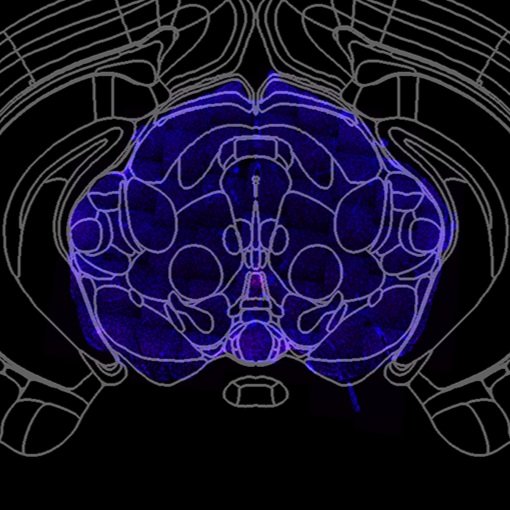
slice-to-atlas registration
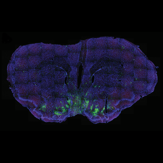
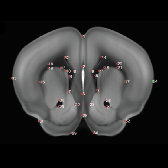
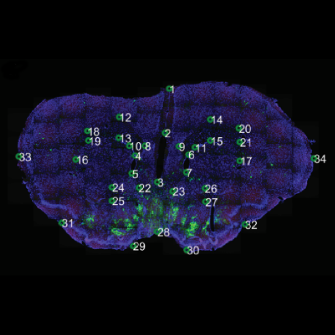
Histological slices are imperfect, making brain regions difficult to accurately delineate. Hand-drawing region borders is a painstakingly slow process. With user-selected landmark points, SHARCQ warps the slice image to match the reference atlas. After this so-called atlas registration is complete, brain region borders are overlaid onto the registered slice.

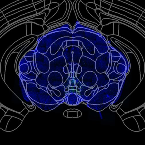
Adjustable Atlas Tilt
During histological sectioning, it is difficult to ensure that all sections are perfectly aligned to the plane of the brain atlas. To address out-of-plane histology, the user can adjust the tilt of the atlas in SHARCQ. The dorsal-ventral and medial-lateral angles can be adjusted independently. This functionality ensures that the atlas registration is accurate across the entire histological section.

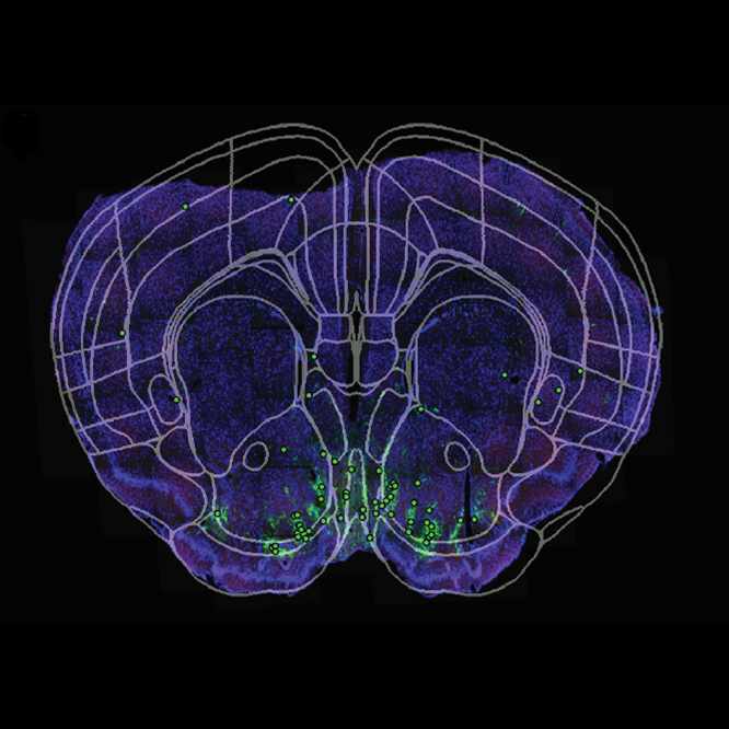
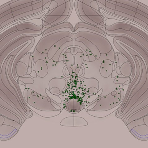
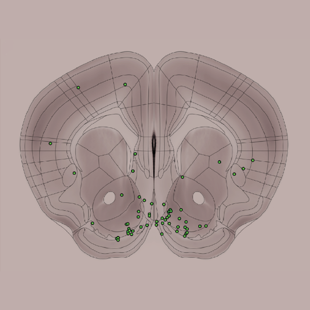
Cell Count Registration
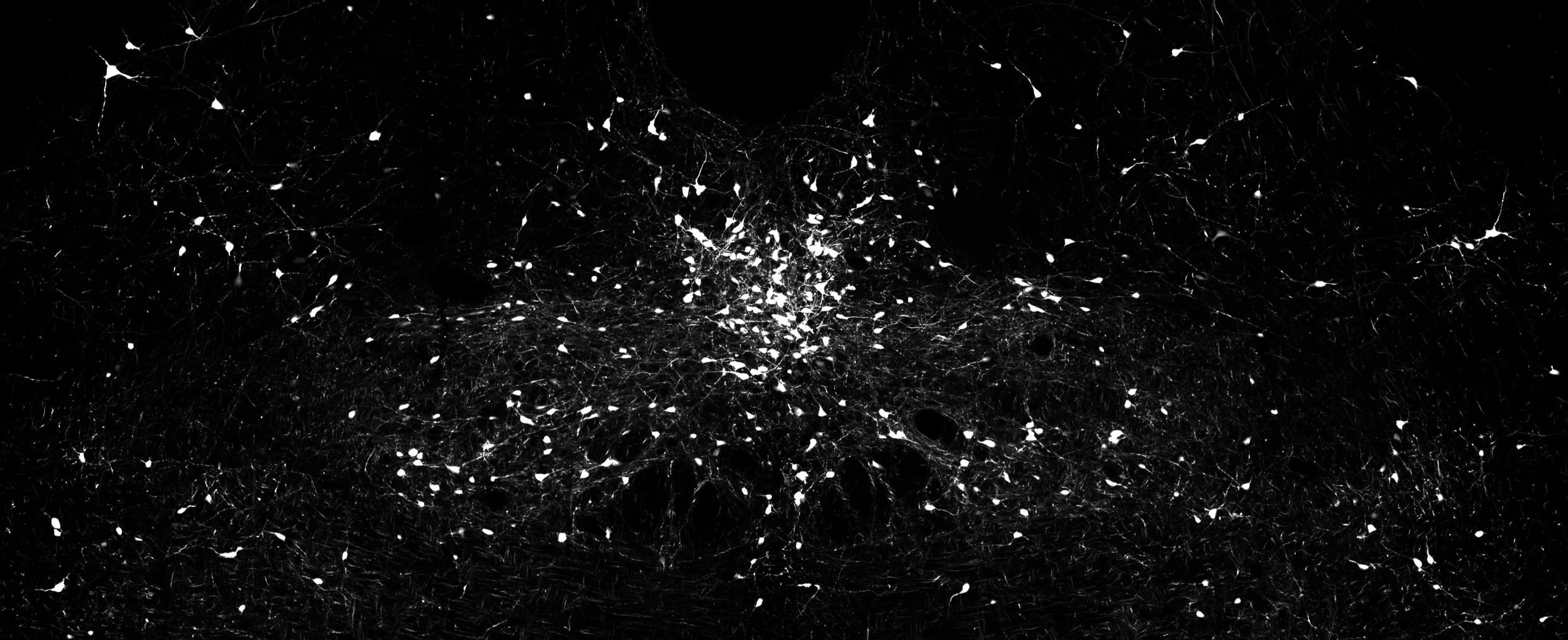
User-inputted cell count files are converted into binary matrices to preserve precise cell locations. The matrix is registered to the atlas using the warping function obtained during slice image registration. Cell quantification by region is also given in tabulated form. SHARCQ accommodates count files from any manual or automatic counting program. Here, each green dot represents one c-Fos-positive cell.

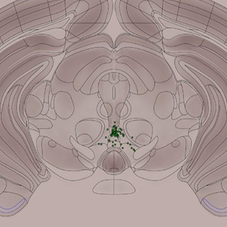
Interactive cell counts
During brain retrieval, excessive distortion of the tissue can occur, such as enlargement of the ventricles. This makes accurate atlas registration very difficult. To circumvent this and other registration issues, all cell locations are fully interactive in SHARCQ. The user can select one cell or draw a polygon to select multiple cells. Selected cells can be moved to the correct region. Cells can be deleted or added in SHARCQ as well.

3D RenderinG
Whole-brain cell counts in serial sections are aligned to generate an interactive 3D wire frame brain. Multiple cell populations can be visualized simultaneously, thereby providing an intuitive snapshot of circuit anatomy between distinct cells. Labeled here are putative glutamatergic neurons in the ventral tegmental area (red) and their direct synaptic inputs across the entire brain (cyan).
Multiple Cell Populations
Cell quantification may take into account multiple discrete populations of cells in the same slice. In these cases, the user performs atlas registration as normal with one count file, then inputs subsequent count files without re-registering. SHARCQ applies an identical warp to all count files to (1) ensure identical region analysis to separate cell populations, and to (2) streamline the workload by requiring the user to perform the registration only once.

Multiple Atlas capability
SHARCQ currently contains data for the Allen Mouse Brain Atlas and the modified Franklin-Paxinos Mouse Brain Atlas. The user can analyze cell locations with the atlas which most accurately reflects their brain regions of interest. SHARCQ is designed to be forward compatible with digitized atlases for any species, and we hope to integrate functionality for other species in the future.
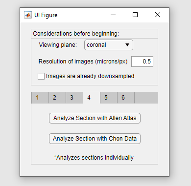
User-friendly GUI
No coding is required to use SHARCQ. The user navigates from step 1 to 6 in the GUI, with additional prompts and controls displayed in the MATLAB command window. SHARCQ was designed to be intuitive and accessible to researchers with little to no coding expertise. Nevertheless, should the user encounter problems, the SHARCQ GitHub contains a troubleshooting guide.